
Providing high-quality echocardiography and vascular duplex imaging.
Call and schedule your appointment today!
718.497.1429
What is diagnostic testing?
Diagnostic testing is used to help your doctor determine the exact condition that is causing your pain or other symptoms. Diagnostic procedures can help your doctor pinpoint your exact underlying problem as well as monitor your progress throughout treatment.
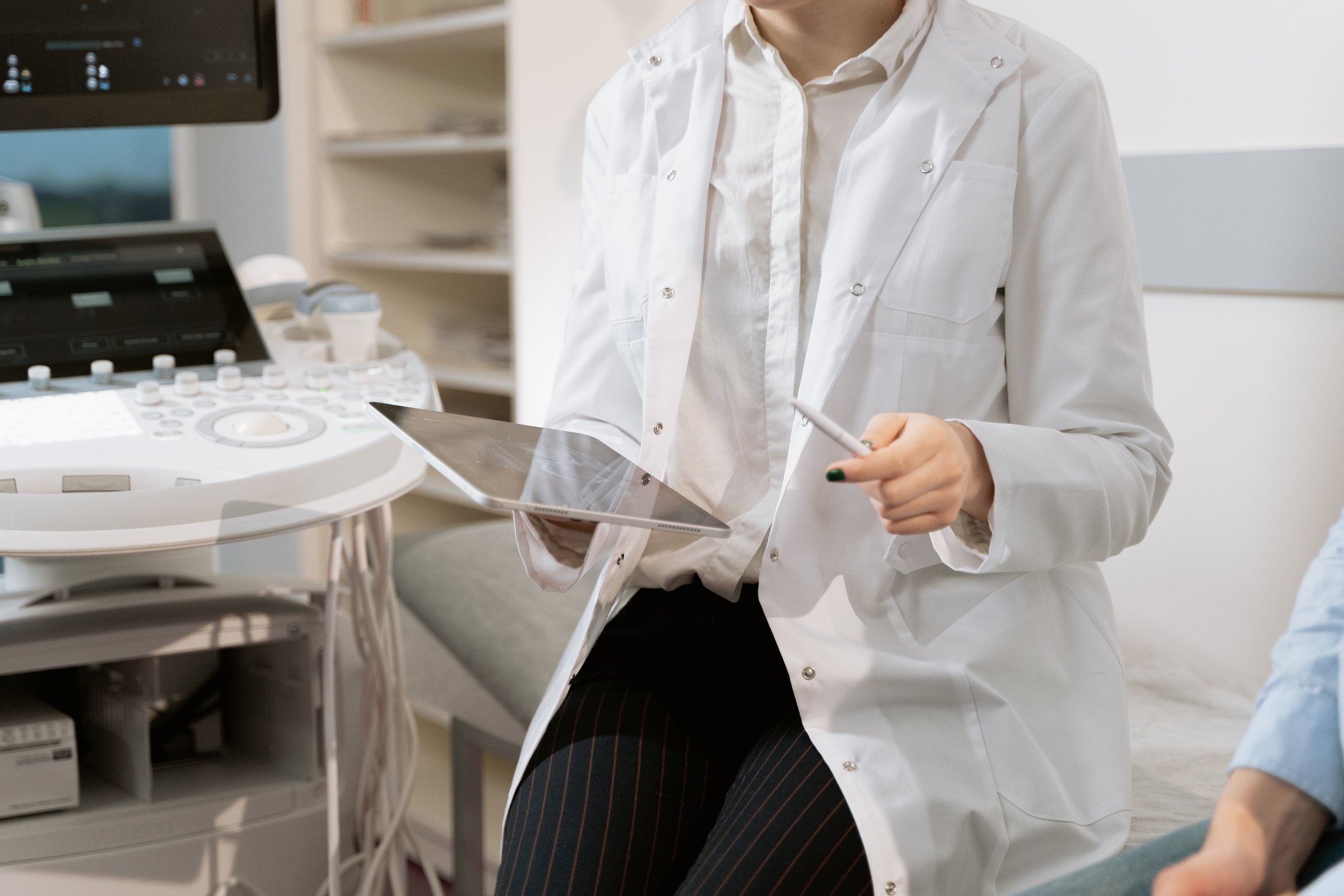
Diagnostic Services
-
Arterial Exam
PURPOSE The upper extremity arterial exam is able to demonstrate blocked or reduced blood flow through the major arteries by detecting stenosis (narrowing), occlusion (complete blockage) or aneurysm (abnormal widening or ballooning).
PROCEDURE The patient lies face up while the ultrasound technologist presses the transducer against the skin at the areas of interest until the desired images are captured.
DURATION Examination time is about 30 minutes
Venous Exam
PURPOSE Imaging of the upper extremity veins is performed to assess the deep and superficial venous system to determine the presence or absence of pathology, and to rule out deep or superficial vein thrombosis.
PROCEDURE The patient lies face up while the ultrasound technologist presses the transducer against the skin at the areas of interest until the desired images are captured.
DURATION Examination time is about 30 minutes
-
Arterial Exam
PURPOSE This procedure provides an overview of the location, extent and severity of vascular diseases, such as claudication (pain upon exertion), rest pain, arterial ulcers, aneurysms (ballooning), and arterial trauma.
PROCEDURE The patient lies face up with the leg externally rotated. The ultrasound technologist then presses the transducer against the skin in various locations along the leg, sweeping over the area of interest until the desired images are captured.
DURATION is about 30 minutes.
Venous Exam
PURPOSE This procedure is performed to assess the venous system of the lower extremities to determine the presence or absence of deep or superficial thrombosis (blood clots).
PROCEDURE The patient lies face up with the leg externally rotated. The ultrasound technologist then presses the transducer against the skin in various locations along the leg, sweeping over the area of interest until the desired images are captured..
DURATION is about 30 minutes.
-
Abdominal Aorta Exam
LOCATION The aorta, which carries oxygenated blood to the lower half of the body and is the largest artery in the body, passes down through the rear of the abdomen to the left of the spine.
PURPOSE The abdominal aorta exam can detect, measure and monitor either a blockage in the aorta or a ballooning of the aorta called an aneurysm. Undiscovered ruptured abdominal aortic aneurysms are the 10th most common cause of death of men in the United States.
PROCEDURE The patient must not drink or eat anything four hours prior to the exam, and is positioned lying face up with the knees slightly bent. The ultrasound technologist then presses the transducer against the abdominal skin, moving back and forth to capture clear images of the blood vessels and arterial structure.
DURATION Examination time is about 30 minutes.
Abdominal Inferior Vena Cava Exam
LOCATION The inferior vena cava (IVC), which carries de-oxygenated blood from the lower body back to the heart, passes up through the rear of the abdomen to the right of the spine.
PURPOSE The IVC exam checks for deep vein thrombosis (clotting), compression, blockage, dilation, and collapse.
PROCEDURE The patient must not drink or eat anything four hours prior to the exam, and is positioned lying face up with the knees slightly bent. The ultrasound technologist then presses the transducer against the abdominal skin, moving back and forth to capture clear images of the blood vessels and venous structure.
DURATION Examination time is about 30 minutes.
-
PFT & Spirometry
-
Sudomotor Neuropathy
Sudomotor dysfunction is a common feature of diabetic autonomic neuropathy. This generally manifests as ( anhidrosis - the inability to sweat normally) or ( hyperhidrosis - excessive sweating.) This is a non- invasive test that is a fast and accurate method for detecting early stages of neuropathies.
-
PURPOSE The goals of a carotid ultrasound exam are to screen patients for blockage or narrowing of their carotid arteries, which, if present, may increase their risk of having a stroke, and to rule out calcium deposits, plaque, and other irregularities. Other risk factors calling for carotid ultrasound are diabetes, elevated blood cholesterol, and a family history of stroke or heart disease.
PROCEDURE The patient is positioned lying face up on the examination table. The ultrasound technologist then presses the transducer against the skin in various locations along the neck, moving over the area of interest until the desired images are captured. It may be necessary to tilt or rotate the head for the best exposure, as the transducer is swept over the entire length of the neck on both sides to obtain views of the arteries from different perspectives.
DURATION Examination time is about 35 minutes.
-
Breast ultrasound is non-invasive and often used as a follow-up test after an abnormal finding on a mammogram, breast MRI or clinical breast exam. If a needle biopsy is needed, breast ultrasound may also be used to help guide the procedure.
-
PURPOSE A renal ultrasound exam is used to detect blockage and abnormalities in the kidneys and urinary tract.
PROCEDURE The patient must not drink or eat anything four hours prior to the exam, and is positioned lying face up with the knees slightly bent. The ultrasound technologist then presses the transducer against the abdominal skin, moving back and forth to capture clear images of the kidneys, renal blood vessels, and urinary tract.
DURATION Examination time is about 30 minutes
-
Ultrasound (Echo, Vascular, General, Small Parts, MSK)
-
EMG, NCV, TCD & VNG
-
PURPOSE Echocardiography is routinely used in the diagnosis, management, and follow-up of patients with any suspected or known heart disease. It is a diagnostic test in cardiology for assessing ischemia (reduced blood flow), valvular dysfunction, regurgitation (backward flow of blood), stenosis (incomplete opening of the heart valves), cardiac tumors and cardiomyopathies (diseases affecting the heart muscle).
PROCEDURE The patient lies face up as wires are placed on the chest to monitor heart rhythm, then turns to lie on the left side as the ultrasound technologist then presses the transducer against the chest in various locations, sweeping over the area of interest until the desired images are captured. The sounds that come from the ultrasound machine are representations of blood flow within the heart.
DURATION Examination time is about 35-45 minutes.
-
PURPOSE When the body is active, it requires more oxygen than when it is at rest, and, therefore, the heart has to pump more blood. Due to the increased stress on the heart, exercise can reveal coronary problems that are not apparent when the body is at rest.
The stress test helps doctors determine how well the heart handles the increased demands imposed by physical activity. It is particularly useful for evaluating possible Coronary Artery Disease, detecting inadequate supply of oxygen-rich blood to the tissues of the heart muscle (Ischemia), and determining safe levels of exercise in people with existing heart disease.
PREPARATION Patients should NOT eat or smoke several hours before the test. The healthcare provider will inform you of taking any of your routine medications on the day of the test.
-
PURPOSE: Instead of looking for heart and coronary artery disease as with adults, Pediatric Echocardiography is used to screen for congenital or acquired heart disease or defects in children.
DURATION: 20-40 minutes. It is important that the child remain still to obtain acceptable high-quality diagnostic images. In some instances, younger children may require sedation to allow the pediatric echo technologist to obtain a complete study.
-
Holter Monitor & 24 Hour BP Monitoring
If you believe you may benefit from diagnostic testing, schedule an evaluation. Our expert physicians can help you determine the underlying cause of your pain and recommend treatment options. Call our Queens office at 718.497.1429